1. Name of the location of 90% of epistaxis
2. A genetic disorder that forms AV malformations in the skin, lungs, brain etc
3. Name of posterior vascular plexus in the nasal cavity causing posterior epistaxis
4. 1st line treatment for all epistaxis
5. The common brand name for anterior nasal packing
6. Chemical used in cautery sticks
7. Physically scaring complication of posterior nasal packing with foleys catheter
Coming soon..
Lower Motor Neurone Facial Palsy
Objectives
1. Know the anatomy and function of the facial nerve and its branches, and be able to describe its intra- and extra-temporal course.
2. Know the incidence/prevalence, clinical presentations, management, and prognosis of the various causes of facial palsy.
Incidence
Annual incidence of 15-30 cases per 100,000 population
Bell's Palsy accounts for approximately 60-75% of cases of acute, unilateral, lower motor neurone facial paralysis.
Bell's palsy is commoner in pregnant women, especially in the third trimester.
Differential diagnosis
There are many methods for formulating a differential diagnosis for facial palsy. One particularly useful one is the surgical sieve. There are various versions of the sieve but the one we use here is VITAMIN D.

Bell's Palsy and Ramsay Hunt Syndrome
Management of these two conditions centres on one simple principle: you must attend to the condition and to the eye.
Clinically, these diseases are quite different but they both cause a lower motor neurone facial palsy and both threaten the eye through its inability to protect itself by blinking.
Bell's Palsy.
This is by far the commonest cause of an acute lower motor neurone (LMN) paralysis and it is a diagnosis of exclusion.
Often, the paralysis arises over night and there may be a viral prodrome. The paralysis then develops quickly to its maximum degree and, over a period of weeks, it usually resolves. However, around 5% of patients will make a poor recovery.
Not all patients have a complete palsy, in fact the majority don't. Instead they have a partial paralysis. This is, nonetheless, alarming for them and they need counselling to understand the natural history of the disease.
Oral steroid is recommended and has an effect in improving the likelihood and degree of recovery. Around 87% of patients will recover fully and the remainder will be left with a partial or, rarely, a complete palsy.
Antiviral therapy is not routinely recommended even though this is considered a herpes simplex virus type 1 infection.
NB. Always attend to the eye. The facial nerve innervates orbicularis oculi and is crucial for normal blinking. The blink protects the eye and syphons tears into the nasolacrimal duct. Thus, patient with a facial palsy may have apparently excessive lacrimation due to a reduced outflow from the eye. This despite the paralysis threatening tear production via the greater superficial petrosal nerve.
Ramsay Hunt syndrome (type 2).
Around 12% of patients with a LMN facial palsy will have Ramsay Hunt syndrome. This is otherwise known as herpes zoster oticus. The key features that separate this condition from Bell's palsy is the presence of a vesicular rash over the pinna, ear canal, face or soft palate. It is also very painful and can leave a neuralgia once the outbreak has gone.
A small number of patients do not show the rash.
Facial paralysis has a much worse prognosis in this condition and it can affect adjacent cranial nerves causing deafness, vertigo and other symptoms.
Treatment is as for Bell's with the addition of acyclovir as long as the disease presents early.
Anatomy
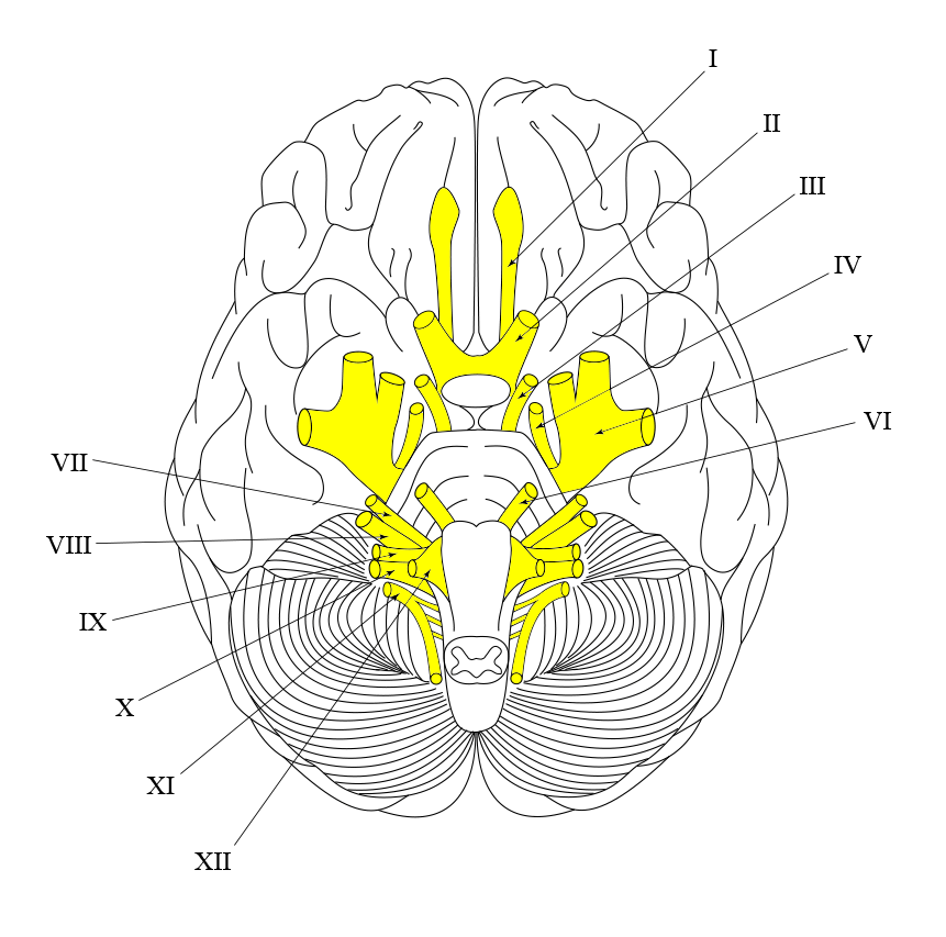
The facial nerve (the 7th cranial nerve or CN VII) originates in the pons of the brain. It forms from two separate nerves; a large motor nerve and smaller sensory root (the intermediate nerve). These nerves fuse to form the facial nerve after passing through the internal auditory meatus and into the facial canal.
The geniculate ganglion is a collection of nerve cell fibres and bodies (Genu (Gk) = knee). It lies antero-superiorly in the medial wall of the middle ear.
The facial canal is a 'Z' shaped structure which encloses the nerve as it passes through the remainder of its intra-temporal path.

Before leaving the skull, the facial nerve gives rise to a number of branches:
Greater superficial petrosal nerve
Synapses with the sphenopalatine ganglion which provides parasympathetic nerve innervation to the lacrimal gland and the mucosal glands of the nose, palate, and pharynx.
Nerve to the stapedius
Innervates the stapedius muscle which helps dampen excessive movement of the stapes.
Chorda Tympani
Special sense of taste from the anterior two-thirds of the tongue.

The facial nerve passes through the stylomastoid foramen, exiting the skull to start its extracranial path.
The nerve passes anteriorly and through the parotid gland where it branches to form five motor branches.
A great mnemonic to remember these branches is
"To Zanzibar By Motor Car"
These nerves innervate the muscles of facial expression as listed below.

- Frontalis, orbicularis oculi and corrugator supercilii
- Innervates the orbicularis oculi
- Innervates the orbicularis oris, buccinator and zygomaticus muscles.
- Innervates the mentalis muscle and other muscles of the lower lip.
- Innervates the platysma
It is only through a thorough knowledge of the underlying anatomy of the facial nerve that a good understanding of the importance of clinical findings and examination can be achieved.
Clinical examination
The first part of the examination is to establish if a facial nerve palsy exists! The level of weakness varies from dense weakness to very subtle. We do this by testing the 5 branches and corresponding facial expression.
Temporal branch

Get the patient to raise their eyebrows.
Zygomatic branch

Ask the patient to close their eyes tightly and keep them closed against resistance.
Buccal branch

Request the patient puffs out their cheeks.
Marginal mandibular branch

Ask the patient to smile showing their teeth.
Cervical branch

Harder to explain without demonstrating yourself. Get the patient to grimace, showing the platysma muscles of the neck.
Upper v Lower motor neurone causes
It is important to be able to distinguish an upper from a lower motor neurone weakness. Remember that muscles of the forehead have bilateral cortical representation.
Let's study the diagram below. Upper motor neurones from the two hemispheres of the cerebral cortex are represented with differing colours. The patient's left cortex are coloured red, the right cortex are represented by yellow.
In the left diagram we see that each facial nerve nuclei (grey circle) receives upper motor neurone input from both sides of the cerebral cortex (via left and right corticobulbar tract in the posterior limb of the internal capsule). As seen by the mixed red and yellow coloured lines, the temporal branch of the face receives innervation from both hemispheres. The remaining branches of the facial nerve, however, only have single hemisphere innervation (single coloured lines).
In the middle diagram there is a lesion of the lower motor neurone (blue circle). This could be a Bell's Palsy, for example. The pathology interrupts the nervous supply of all of the facial nerve taking out both the yellow (right) and red (left) cortical nervous innervation to the facial muscles. As such there is ipsilateral weakness in all of the facial divisions on the right side of the face (pale blue coloured side).
Therefore, in lower motor neurone pathology, we expect all branches of the facial nerve to be weakened.
In the right-most diagram the red circle represents an upper motor neurone (UMN) pathology in the left side of the patient's brain e.g. a stroke. We would expect, from our neurology anatomy, the right side of the patient's face to be weakened (dark pink side). The stroke disrupts the red nervous innervation to the face (left corticobulbar tract), however the right side of the patient's brain (yellow) still gives innervation to the upper branches of the facial nerve, through the pathway described above.
This means that the forehead or temporal branch of the facial nerve is spared. So, we can deduce an UMN lesion clinically in a facial palsy by the sparing of the forehead muscles from weakness.

The House-Brackmann classification
It is important to be aware of the method used for classifying the severity of a facial weakness. The table below outlines this classification. There is no need to memorise this, just be aware that it exists.

Other areas to examine
A full and thorough examination must be conducted to ensure no pathology is missed and the correct diagnosis is made. Areas to specifically focus on are the parotid, the ear, oral examination, and a thorough neurological examination including cranial nerves.
If a lower motor neurone palsy does not resolve within three weeks the patient is usually referred to ENT and a baseline audiogram is performed to help rule out an acoustic neuroma. An MRI scan of the brain will be considered if there is no recovery at all.
Below is an example of a commonly used facial palsy sticker that is completed and documented in the notes. It summarises examination and treatment nicely.


