1. Name of the location of 90% of epistaxis
2. A genetic disorder that forms AV malformations in the skin, lungs, brain etc
3. Name of posterior vascular plexus in the nasal cavity causing posterior epistaxis
4. 1st line treatment for all epistaxis
5. The common brand name for anterior nasal packing
6. Chemical used in cautery sticks
7. Physically scaring complication of posterior nasal packing with foleys catheter
Coming soon..
Inner Ear Anatomy
Remit
This page gives you the fundamentals of the anatomy of the vestibular portion of the inner ear. It is important for you to know this before you try to understand vestibular physiology.
Anatomy
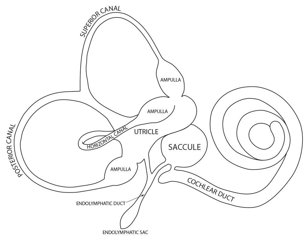
The inner ear is a complex three dimensional shape with semicircular canals, dilations called the utricle and saccule and a spiral portion known as the cochlea. All of these organs are housed inside a bony shell known as the bony labyrinth and this is within the temporal bone. The cochlea is the site where sound is transformed into neural energy for hearing. The rest is concerned with balance.

The Right Membranous Labyrinth
This diagram shows the membranous labyrinth and it is this that contains the neuroepithelia that detect motion and sound.
The bony and membranous labyrinths are separated by a fluid filled space and this is shown diagrammatically below.
Despite the complexity of it's shape it can be simplified as in the following diagram.

The yellow central portion in the diagram represents the membranous labyrinth. It contains endolymph and all of the neuroepithelia required for hearing and balance. Surrounding this membranous labyrinth is a fluid filled space. This space separates the membranous labyrinth from the bony labyrinth. The space is filled with perilymph and can be considered to act in the same way as CSF - as a cushion for the delicate structures it protects.
The endolymph is produced by 'Dark Cells' within the membranous labyrinth. The dilated portion in this diagram represents the endolymphatic sac (ES). This is thought to regulate the volume and composition of the endolymph although how this is done is open to speculation.
If the composition or specific gravity of the endolymph is changed as in Ménière's Syndrome or alcohol consumption, the function of the balance and hearing epithelia within it are affected. Both Ménière's and Alcohol intoxication are explained in other tutorials.
The Right Membranous Labyrinth
The inner ear is supplied by the Superior Vestibular Nerve, the Inferior Vestibular Nerve and the Cochlear Nerve. All of these nerves travel from the inner ear towards the brain stem within the Internal Acoustic Meatus. Along with these in the IAM is the Facial Nerve and the vascular supply.
The Right Membranous Labyrinth - Nerve supply
In this diagram we visualise the inner ear. The cochlea nerve comes from the cochlea but that is not shown.
The utricle, some of the saccule, the lateral semicircular canal and the superior semicircular canal are all supply the superior vestibular nerve. This part of the labyrinth is sometimes called the superior labyrinth because of how it develops embryologically.
The posterior canal supplies the inferior vestibular nerve. This nerve is also supplied by part of the saccule. This part of the labyrinth is sometimes called the inferior labyrinth.

So the superior vestibular nerve takes its input from the superior labyrinth (LSCC, SSCC, and utricle) and then passes out along the internal acoustic meatus to the vestibular nucleus on the same side.
The inferior vestibular nerve takes its input from the inferior labyrinth (PSCC and the saccule). It joins with the superior nerve and travels to the vestibular nucleus too.
The Right Membranous Labyrinth - Blood supply
The diagram below outlines the blood supply for the inner ear. The anterior vestibular artery supplies the utricle, superior canal and the lateral canal (superior labyrinth). The posterior vestibular artery supplies the posterior canal (inferior labyrinth). Both of these are branches of the labyrinthine cochlear artery which is derived from the anterior inferior cerebellar artery.

An end artery system
Many areas of the body have a dual blood supply. For example the hand gets blood from two arteries; the radial and the ulnar. The brain gets blood from even more. It has four major arteries. The means that these structures are protected when one artery becomes blocked because blood can get in from another direction.
The diagram shows that the blood supply to the inner ear comes from only one source, the labyrinthine artery. This means that the inner ear structures cannot get blood from anywhere else if there is a blockage caused by a clot or a thrombosis. This is known as an end artery system and it makes the inner ear vulnerable to damage.
The Right Internal Acoustic Meatus (IAM)
The IAM is the bony conduit in the petrous temporal bone through which the vestibular, cochlear and facial nerves travel. Note that there are two vestibular nerves on each side, a larger superior and a smaller inferior nerve. The diagram shows the right IAM seen from the pons.

The Right Internal Acoustic Meatus
In the diagram above imagine standing at the brainstem and looking outwards towards the Right ear.
S= superior, I= inferior, A= anterior, P= posterior.
VII is the Facial Nerve, C is the Cochlear Nerve,
SV is the Superior Vestibular Nerve, and IV is the Inferior Vestibular Nerve.
The vertical line represents Bill's Bar while the horizontal line is the Crista Falciformis. The Nervus Intermedius travels with the Facial Nerve and is not shown.
Strictly speaking the two vestibular nerves and the cochlear nerve are travelling towards the brainstem in the IAM and the facial nerve is travelling away from the brainstem. This is because nerves should be described by the the direction of the nerve impulses in them. The ear generates the impulses and sends them to the brain.
The Vestibular Nuclei
The vestibular nerves travel towards the brainstem through the internal acoustic meatus and enter the brainstem at the pons. The nerve fibres synapse with cell bodies in the vestibular nuclei. These nuclei lie in the floor of the fourth ventricle.


The left image shows the brain stem and cerebellum viewed from the side. The vestibular nuclei are mostly in the pons but they extend down into the medulla a little. The right image shows a view of the brainstem with the cerebellum removed. The vestibular nucleus can be seen in the floor of the fourth ventricle.
The vestibular nucleus consists of four smaller nuclei: the superior, medial, lateral, and descending nuclei.
The superior and medial nuclei are mostly involved with the vestibulo-ocular reflex and send nerve fibres up the brainstem towards the nuclei that are involved with eye movement: the oculomotor, abducent and trochlear nuclei. These nerve relays travel in the medial longitudinal fasciculus.
The lateral vestibular nucleus is involved with vestibular-spinal reflexes and has an outflow that descends towards the neck and the rest of the spinal cord.
The commissural system
The vestibular nuclei on the left are joined to the ones on the right by a system of nerve fibres that cross the midline. This is the commissural system and it is inhibitory in nature. This means that the right vestibular nuclei inhibit the activity in the left nuclei and vice versa. This is important in the push-pull system related to the functional pairs of the semicircular canals.
A functioning inhibitory commissural system is vital for the proper management of eye position during gaze and damage to the system or loss of the proper balance of activity in the system leads to abnormalities such as gaze-evoked nystagmus.
Other inputs
The vestibular nuclei also receive sensory information from the eyes, somatosensory fibres from pressure receptors especially in the feet, and proprioceptive fibres from joints. Thus they are far more than simple nuclei for information from the ears.

