1. Name of the location of 90% of epistaxis
2. A genetic disorder that forms AV malformations in the skin, lungs, brain etc
3. Name of posterior vascular plexus in the nasal cavity causing posterior epistaxis
4. 1st line treatment for all epistaxis
5. The common brand name for anterior nasal packing
6. Chemical used in cautery sticks
7. Physically scaring complication of posterior nasal packing with foleys catheter
Coming soon..

Pictures of Diseased Ears
On this page you will find clinical images of diseased ears with a description of the abnormally / disease present.


Above. Left ear with a central, inferior, perforation. This ear had very little hearing loss and the ossicular chain was intact. Some of the perforation is hidden in the anterior recess of the ear canal. This recess can be very deep, making a view of all of the drum difficult.


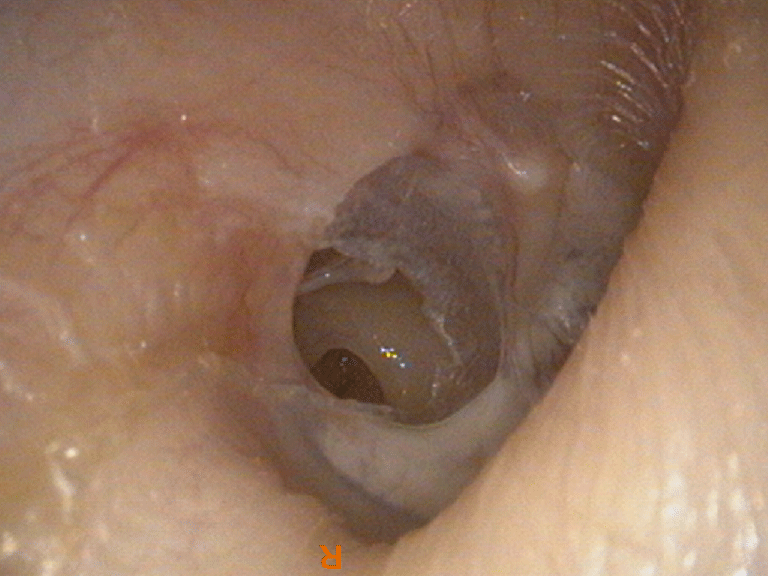
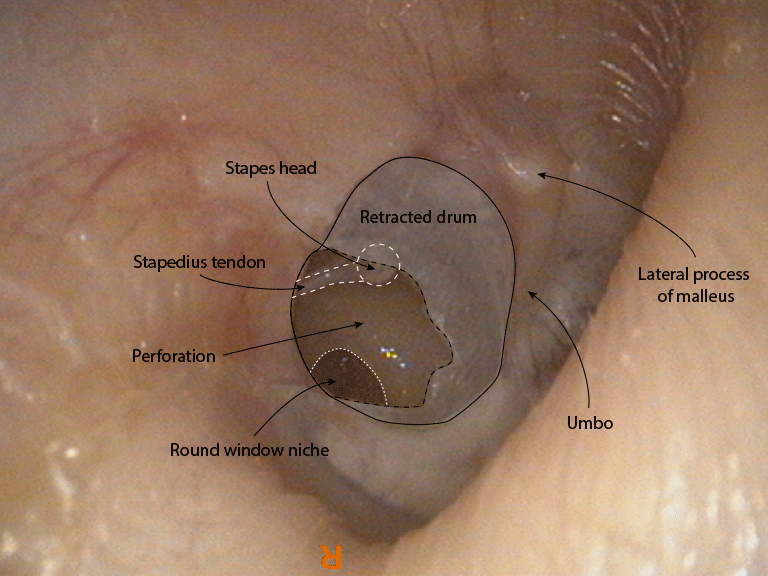
Above. Right ear with a posterior, marginal perforation. In this ear the posterior drum became weakened and retracted onto the long process of the incus and stapes head. With time the long process dissolved away leaving the head intact. At some point he drum ruptured and a perforation appeared. This patient had a significant conductive hearing loss due to the loss of the continuity in the ossicular chain and because the round window had become exposed by the perforation.


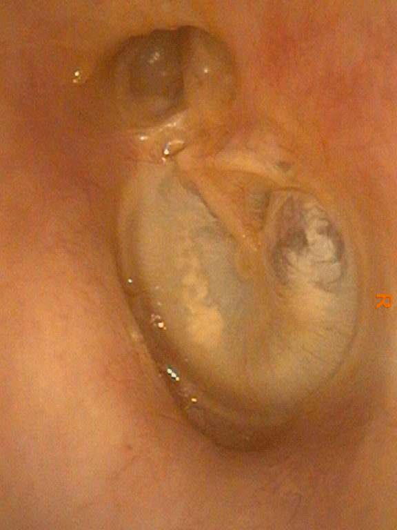
Above. A left ear with an attic perforation. This small hole led into two sacs of skin. Only one is shown in the diagram, lying medial to the anterior half of the eardrum. This ear had a recurring scanty discharge and a foul smell. There was a conductive hearing loss because the body of the incus had been eroded.
Surgery removed the disease but had to remove the incus and the head of the malleus too because the second sac had wrapped itself around both.
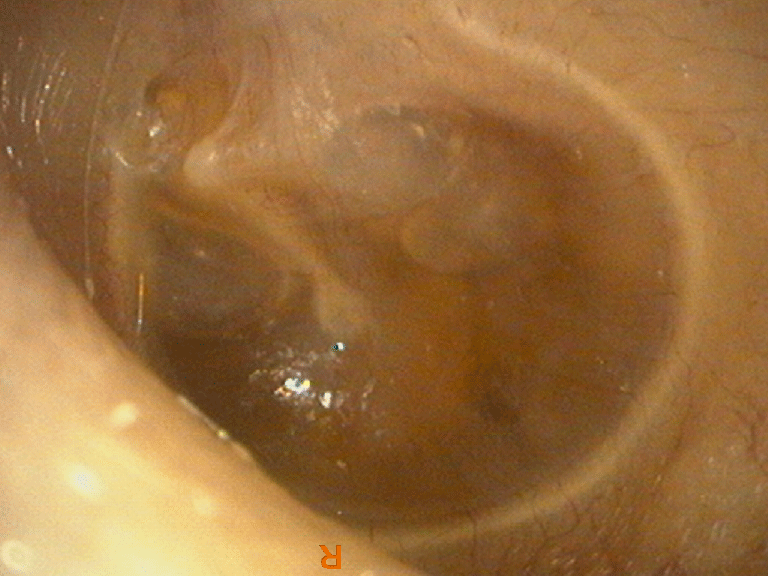
Above. Another right ear with a posterior marginal perforation.

Above. A left ear with an attic retraction pocket. The pocket was wrapped around the anterior side of the malleus head. It wasn't very deep but it did become infected often and this is what took the patient to his doctor. Surgery was performed via an endoscopic approach with complete removal of disease and without disruption of the ossicular chain. Normal hearing was preserved.

Above. A left ear with a middle ear effusion. The effusion appears amber / honey in colour but can look grey and dull. This one has a number of air bubbles within the fluid. This is usually taken to mean that the middle ear effusion is resolving and air is getting up the Eustachian tube in to the middle ear.
Above. A left ear with a middle ear effusion. The effusion appears amber / honey in colour but can look grey and dull. This one has a number of air bubbles within the fluid. This is usually taken to mean that the middle ear effusion is resolving and air is getting up the Eustachian tube in to the middle ear.

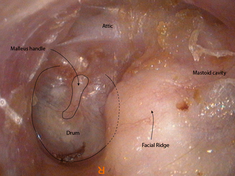
Above. A left ear with a modified radical mastoid cavity. The outer attic wall (scutum) has been removed and the malleus head and incus have been removed top clear disease. They would normally be in the attic space. The mastoid air cells have also been opened to remove disease. The facial nerve lies somewhere under the 'facial ridge'. This ridge is not an anatomical structure. It is created during surgery.
Cavities like this are fast being replaced with more modern approaches but they still have their uses.
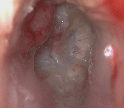
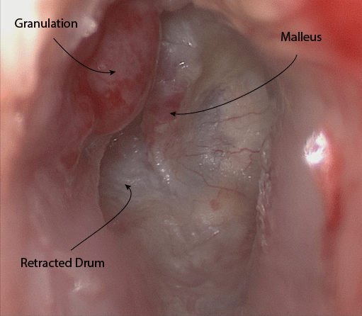
Above. A right ear with a retracted posterior half and granulations on the bone adjacent to the retraction. These granulations mean that there is infection present and it is probably arising from cholesteatoma. The ear is likely to have a foul smell. If it is possible a CT scan prior to surgery is required by most modern surgeons.