1. Name of the location of 90% of epistaxis
2. A genetic disorder that forms AV malformations in the skin, lungs, brain etc
3. Name of posterior vascular plexus in the nasal cavity causing posterior epistaxis
4. 1st line treatment for all epistaxis
5. The common brand name for anterior nasal packing
6. Chemical used in cautery sticks
7. Physically scaring complication of posterior nasal packing with foleys catheter
Coming soon..

Interpreting CTs of the Temporal Bone
Studying CT scans can help with your understanding of anatomy and is a vital prelude to understanding pathological findings revealed by the investigation. On this page are presented normal CT anatomy in two planes and a selection of pathological scans with commentary.
There are some things that you need to know before you start.
Key facts.
-
As with CT scans of the paranasal sinuses, abnormalities in the ear are common, especially the presence of fluid in the mastoid air cells.
-
Using a CT to diagnose problems is, therefore, a little unreliable by itself. They should only be organised after a careful otology history and examination.
-
A CT scan is used when we are planning surgery as it gives a good view of the anatomy of the ear. It can sometimes help show anatomical variations that might cause risk to the patient during surgery. They are commonly requested when a complication of ear disease has arisen, such as facial palsy or labyrinthitis.
-
Plain X-rays of the ears were commonly done in the past but CT and MRI have superseded these.
Introduction
A good knowledge of the anatomy of the ear is essential before trying to understand the CT anatomy. CT scans can be formatted in any plane: axial, coronal and parasagittal. In practice we use the coronal scans most but when we operate on the ear all views are useful. The parasagittal scan is the one that gives the nearest to the surgeon's view during endoscopic surgery. Review the tutorial on anatomy of the ear before proceeding.
Normal Radiological Anatomy
The following sequence of slides shows normal anatomy in the axial plane There are coronal and parasagittal scans too. Once you understand the radiological appearances of the ear we will look at pathology.
Axial Anatomy

Axial CT scan showing the posterior semicircular canal, the IAM, cochlea, and the malleus and incus.
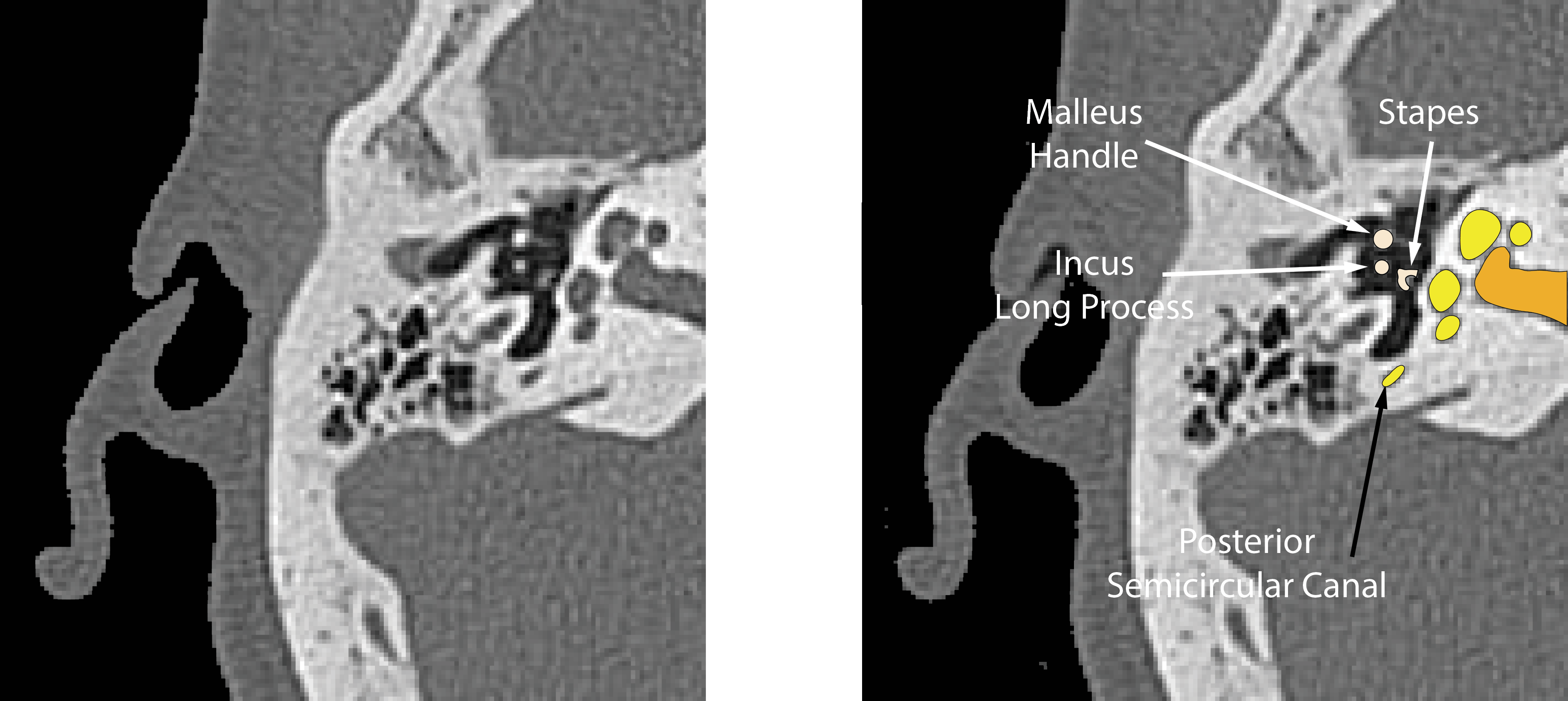
Axial CT scan showing the stapes suprastructure. The malleus handle and the long process of incus are also clearly seen.
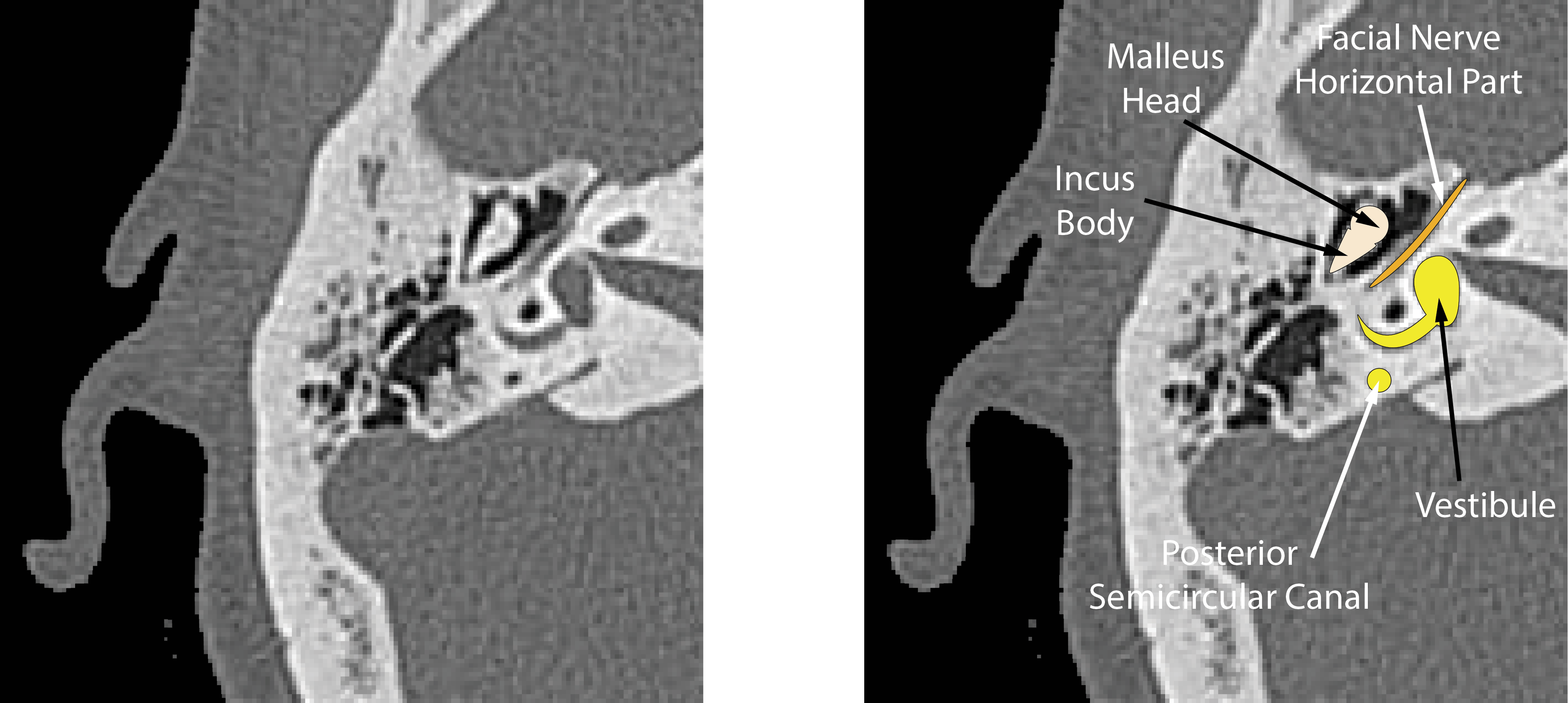
Axial CT scan showing the 'icecream cone' - the malleus head and incus body. The horizontal part of the facial nerve is also visible passing from anterior to posterior.
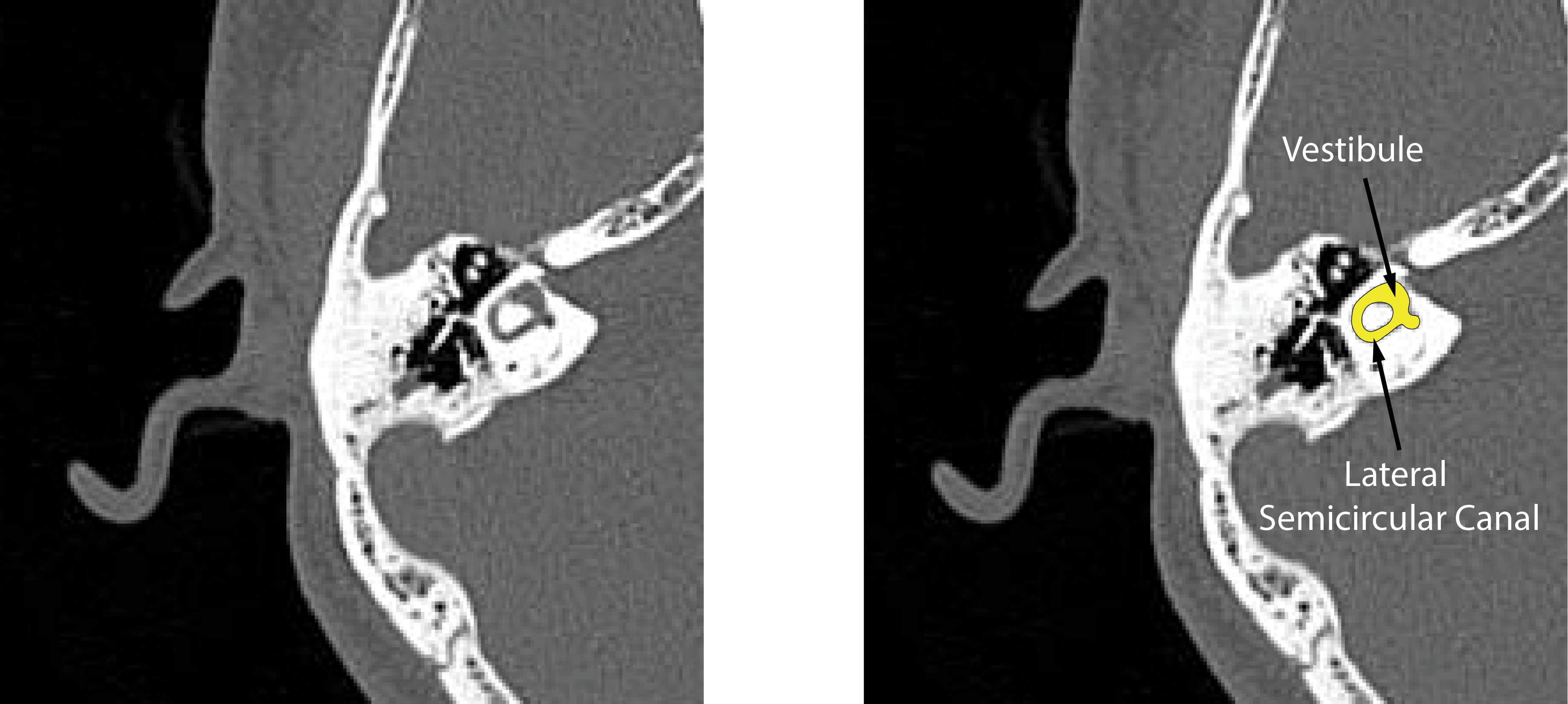
Axial CT scan showing lateral semicircular canal and the vestibule of a different patient. This is the 'signet ring'.

Axial CT through superior and posterior semicircular canals. Air in the epitympanum and mastoid is clearly visible too.
Parasagittal Anatomy

Parasagittal CT through lateral semicircular canal showing the vertical portion of the facial nerve as it runs towards the stylomastoid foramen.
Coronal Anatomy

Coronal CT showing the head and lateral process of the malleus, the ear drum and the cochlea.
Cases with Pathology
Below are examples of CT scans with pathology. It is not usually possible to state what the exact pathology is by looking at a CT scan and clinical examination with histological study give a better understanding. Don’t be tempted to make a diagnosis on the scan alone, always correlate the scan with clinical findings.

Case courtesy of A.Prof Frank Gaillard
This coronal CT shows a cholesteatoma in the epitympanum of the left ear. The lateral semicircular canal is intact but the disease envelopes the malleus. The right ear is normal.

Case courtesy of Prof Frank Gaillard
This right ear shows disease in the epitympanum that extends up to the level of the tegmen. It was found to be cholesteatoma. The disease envelopes the malleus head.

Case courtesy of Dr Andrew Dixon
This coronal CT shows an opacity in the inferior part of the middle ear and attached to the promontory. Other scanning and clinical examination confirmed that it was a glomus tympanicum.

Case courtesy of Dr Eid Kakish
This coronal CT shows a soft tissue mass in the external ear canal. It has widened the canal and is pushing against the eardrum. Air is seen in the middle ear a d the malleus is visible.
This was a canal cholesteatoma.