1. Name of the location of 90% of epistaxis
2. A genetic disorder that forms AV malformations in the skin, lungs, brain etc
3. Name of posterior vascular plexus in the nasal cavity causing posterior epistaxis
4. 1st line treatment for all epistaxis
5. The common brand name for anterior nasal packing
6. Chemical used in cautery sticks
7. Physically scaring complication of posterior nasal packing with foleys catheter
Coming soon..

Benign Paroxysmal Positional Vertigo (BPPV)
Remit
This page describes BPPV, the commonest of vertigo caused by inner ear disease. It is a surprisingly important disease as not only is it the most common cause of vertigo, it is also the most curable.
For an in-depth treatment read: BMJ 1995;311:489 (19 August 1995). Fortnightly review: Benign positional vertigo: recognition and treatment. Lempert, T., Gresty, MA., Bronstein, AM.
Background
There are three main causes of vertigo: Benign Paroxysmal Positional Vertigo (BPPV), Vestibular neuritis and Ménière's disease. They all provoke a spinning sensation and are caused by inner ear disease but that is about all they have in common.
In BPPV the vertigo lasts for seconds only and is provoked by specific movements. It is a disease that most commonly starts without warning (idiopathic) but it also commonly follows head trauma. Interestingly, it may also follow other causes of vertigo - Ménière's Disease and Vestibular Neuritis.
Idiopathic BPPV arises in the 50s and 60s.
It is a condition that remits and relapses. Although the spinning only lasts for seconds the patient will describe periods when it is present most days and periods when it is absent. It affects men and women equally.
Patients describe their vertigo as brief and provoked by: rolling over in bed, looking up or down and bending over and straightening up. These are typical provocations. There is no hearing loss or tinnitus associated with the disease (but beware that these symptoms are common in the general population and may co-exist).
Rule of thumb: Patients who complain that they become dizzy in bed when they roll over to the right usually have right posterior canal BPPV. Likewise, if it is to the left, it is the left posterior semi-circular canal.
Physiology
You should review the section on vestibular physiology before considering the pathophysiology of this disease. In particular you should note the physiology of the macula and cristae and the generation of nystagmus.
Pathophysiology
In BPPV the otoconia attached to the macula of the Utricle become displaced into the endolymph. While floating in the endolymph of the utricle they cause no problems but when they become trapped in the semi-circular canals they induce nystagmus and vertigo of short duration following turns of the head or body.
Classically they become lodged in the posterior semi-circular canal. They do this because of the geometry of the canal. When the patient lies down at night gravity pulls the free floating otoconia into the mouth of the PSCC. On sitting up the following morning gravity pulls them further into the canal until they become lodged behind the cupula in the ampulla.
The posterior canal is vertically orientated so the otoconia cannot escape once they are in behind the cupula.
In the diagram the otoconia are coded as blue.
The posterior canal is by far the most frequently affected (over 94% of cases of BPPV). You can see that if the otoconia were to drift into the canal of the superior canal they would simply fall out again when the patient sat up.
The posterior canal is wired up to the superior oblique and the inferior rectus muscles of the orbit. These are the green coded muscles in the physiology tutorial. Overactivity in the canal causes a characteristic nystagmus that is predominantly rotatory.

Clinical Examination
The history will nearly always point to BPPV as a cause because it is so characteristic in duration and provocation. However, in BPPV there is a sensitive and specific clinical test as well - the Dix-Hallpike test.
The Dix-Hallpike test stimulates the posterior semi-circular canal. You should ask for it to be demonstrated when you do clinical placements. This site shows it in action and there is a video available at the bottom of the page.
The nystagmus is very characteristic. On lying down there is a pause when nothing happens (latency), then there is a geotropic rotatory (or torsional) nystagmus which lasts a 20 -30 seconds, this is followed by rotatory nystagmus in the opposite direction after sitting the patient up (reversibility). If the clinician lies the patient down again the nystagmus is often much reduced or absent (fatigable).
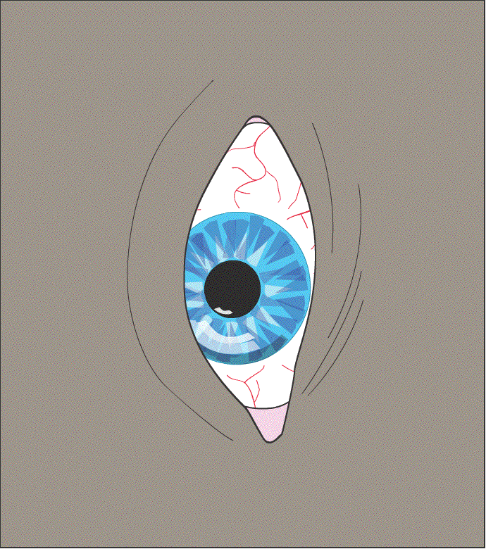
The animated eyeball show the sort of thing to look for. It is best to look at a blood vessel to appreciate what is happening. There is a slow rotation of the eyeball in one direction followed by a quick recovery turn in the opposite direction.
The eye movements of a positive, right-sided Dix-Hallpike test are shown. Remember that in the animation we are looking at the left eye and that the right ear is being tested. The 12 'o' clock position on the eyeball is moving quickly towards the ground. The video of Dix-Hallpike testing below shows the nystagmus.
Treatment
Once diagnosed the patient can generally be cured by an Epley Manoeuvre. This works about 85% of the time and is a simple bedside treatment. There are modifications of this available and other treatments such as Brandt-Daroff and Semont manoeuvres.
Youtube supply plenty of videos of both the Dix-Hallpike and the Epley Manoeuvre. Below are exemplary versions for Swansea University.
Dix Hallpike
Epley Manoeuvre
Do a self-assessment on BPPV here.